Reports on Plant Diseases |
RPD No. 210 - Physoderma Brown Spot of Corn
|
May 1993
|
[ Symptoms ] [ Disease
Development ] [ Control ]
|
Physoderma brown spot disease of corn and of the closely related teosinte
(Zea mays subsp. mexicana) is caused by the fungus Physoderma maydis (P.
zeae-maydis). Brown spot occurs primarily in the southeastern United States,
the Gulf Coast, and the lower Mississippi Valley where yield reductions
from loss of grain and lodging of 25 percent or more have been recorded.
Brown spot is usually a minor disease in Illinois, restricted by weather
conditions to the warmest and most humid regions of the state. The disease
has appeared only occasionally in Illinois since first being found in
1911. A localized outbreak of the disease occurred in southeastern Illinois
in 1971 after considerable rainfall during the early growth of the corn
crop. In a number of bottomland fields near the Wabash and Ohio Rivers,
80 percent or more of the corn plants lodged because of extensive infection
at the nodes under the leaf sheaths, greatly weakening the stalks. Yield
losses caused by the lodging of infected plants increased further during
windy weather as the crop matured.
|
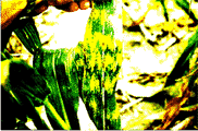
Figure
1. Symptoms of Physoderma brown spot on corn
leaf blades. Yellow halos darken as the corn plant matures.
|
|
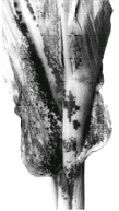
Figure
2. Physoderma
maydis infection of a corn leaf and leaf sheath. Under favorable conditions,
severe stalk rot will develop at nodes beneath the leaf sheath.
|
Symptoms
Brown spot lesions first appear as very small, round-to-oblong, yellowish
spots on the leaf blade (Figure 1), leaf sheath (Figure 2), stalk, and
rarely on the husks and tassel of the outer ear. The spots may occur in
bands across the leaf blade. Infected tissues turn a chocolate brown to
reddish brown and merge to form large blotches with an irregular, angular
appearance (Figure 3). Cells of infected corn tissue disintegrate to expose
dusty pustules (brown blisters) containing enormous numbers of microscopic
sporangia (18 to 24 by 30 to 30 microns). The sporangia are a golden brown
to dark brown. Infection at the nodes beneath the leaf sheaths and the
premature death of plants from leaf blights favor stalk rot and lodging
(Figure 4).
Back to Top
|
|
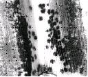
Figure 3. Close-up
of a corn leaf blade showing the chocolate brown blotches, an advanced
stage of Physoderma brown spot. Infected corn tissues contain large numbers
of sporangia that may be released as the corn leaf ruptures and dies.
Brown spot symptoms are most prominent in the leaf midrib area.
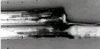
Figure 4. Severe
stalk rotting and lodging may occur when Physoderma maydis invades the
nodes of susceptible corn hybrids. (Courtesy G.L. Scheifele)
|
The thick-walled, brown sporangia (resting spores) formed within infected
cells enable P. maydis to overseason in corn debris or in the soil. The
sporangia are released from infection pustules, disintegrating corn debris,
and soil and are carried to susceptible plants by air currents, insects,
splashing rain or flowing water, and humans. Corn becomes increasingly
susceptible to Physoderma until the plants are about 45 to 50 days old;
susceptibility declines steadily after that.
Free water is required for infection. When moisture is present in the
whorl or behind the leaf sheaths and temperatures are relatively high
(73 to 90 F, 23 to 30 C), a sporangium "germinates" to release
20 to 50 swimming zoospores. The zoospores move about in water for 1 to
2 hours before settling down, becoming amoeba-like, and penetrating yong
meristematic tissue with fine infection hyphae. The resulting mycelium
enters mesophyll or parenchyma cells and forms larger, vegetative structures
(Figure 5). Infection commonly occurs in a diurnal cycle, resulting in
alternating lateral bands of infected and healthy leaf tissue as it emerges
from the whorl. Zoospores of P. maydis can infect corn tissue only during
certain hours of the day and within a few hours after being released.
The development of symptoms and the germination of new sporangia occur
approximately 6 to 20 days after infection, completing the disease cycle
(Figure 5).
Back to Top
|
|
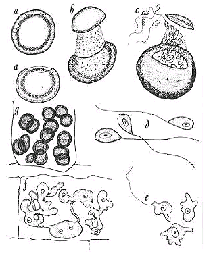
Figure 5. Stages
in the life cycle of Physoderma maydis as seen through a high-power microscope.
Stages a through g can occur in as short a period as 16 to 20 days. (a
Two sporangia (resting spores), top view and side view. (b) stage in opening
of a sporangium, showing the early stage of zoospore formation. Note the
dehisced operculum (lid) being carried up by the enlarging sporangium.
(c) Mature zoospores escaping through the ruptured apex of the resting
spore. (d) Three zoospores with a single flagellum from a germinated resting
spore. (e) Germinating zoospores, amoeboid stage. (f) Rhizomycelium within
a corn epidermal cell showing young sporangia at the ends of short hyphae.
(g) Corn leaf cell filled with mature resting sporangia (Drawing by Lenore
Gray).
|
|
1. Plant resistant hybrids and varieties adapted to your area.
2. Shred infected corn debris in the fall and chisel plow. Tillage may be
done in the fall or spring, depending on the conservation tillage practices
recommended for the area.
3. Avoid planting known susceptible varieties in river-bottom soils or other
high-humidity locations.
4. Rotate crops, since this may contribute to a reduction of inoculum. Physoderma
is known to remain viable in the soil, as sporangia, for about 3 years. Spore-trapping
data indicate that the sporangia can be blown or carried long distances and
be spread easily to adjacent fields. Infected plants transported for silage
can also move considerable numbers of sporangia into disease-free areas.
Back to Top
For further information concerning
diseases of crucifers and other vegetables, contact Mohammad Babadoost, Extension
Specialist in Fruit and Vegetable Diseases, Department of Crop Sciences, University
of Illinois at Urbana-Champaign. University
of Illinois Extension provides equal opportunities in programs and employment.
|